1. Overview of Human Development
Logical starting point
- Human development begins at fertilisation, when a sperm fertilises an ovum.
- Once fertilisation occurs, a zygote is formed.
Definition of an embryo
- An embryo is defined as the organism from the moment mitosis of the zygote begins.
- Therefore:
- Even a 2-cell stage organism is already considered an embryo.
Growth timeline
- Over the first 8 weeks, these few cells:
- Multiply rapidly
- Differentiate
- Organise into tissues and organs
- By the end of 8 weeks, the embryo contains many millions of cells.
- After this point, it is termed a fetus.
Critical vulnerability period
- The embryonic period (weeks 2–8) is the most critical phase.
- During this time:
- Major organs and body systems form
- The embryo is highly vulnerable to:
- Viruses
- Drugs
- Other teratogens
- This is the period when malformations are most likely to occur.
2. Prenatal Stages of Development (Table 11.1 Explained Logically)
Stage 1: Pre-embryonic period (Conception → Week 2)
What happens first and why
- Fertilised ovum undergoes rapid mitotic divisions
- Key events:
- Formation of morula
- Formation of blastocyst
- Implantation of blastocyst
- Development of germ layers
Stage 2: Embryonic period (Week 2 → Week 8)
Structural blueprint phase
- Development of:
- Germ layers
- Placenta
- Formation of:
- All major body systems
Stage 3: Fetal period (Week 9 → Birth)
Growth and maturation phase
- Organs already formed now:
- Grow
- Mature
- Become functional
- Locomotor system becomes functional
3. Gametogenesis – General Concept
What gametogenesis means
- Gametogenesis = formation of definitive germ cells
- Two parallel processes:
- Oogenesis → female
- Spermatogenesis → male
What changes occur
- Both processes involve:
- Cytoplasmic changes
- Chromosomal changes
- Goal:
- Formation of haploid gametes (oocyte or spermatozoon)
4. Key Differences Between Oogenesis and Spermatogenesis
Timing and pattern
- Oogenesis
- Begins in fetal life
- Is cyclical
- Produces usually one oocyte per month
- Spermatogenesis
- Begins at puberty
- Is continuous
- Continues throughout adult life
Hormonal and uterine coordination (female)
- Female monthly cycle includes:
- Oocyte maturation
- Cyclic hormonal changes
- Concurrent endometrial changes
- Purpose:
- Prepare uterus for possible pregnancy
5. Origin and Migration of Primordial Germ Cells
Origin
- Primordial germ cells arise from:
- Wall of the yolk sac
- During the second week of development
Migration
- By the sixth week:
- They migrate into the embryo
- Settle in the gonadal ridges
Proliferation
- Once in gonadal ridges:
- Undergo rapid mitotic divisions
- Proliferation pattern differs between sexes
6. Germ Cell Development – Female vs Male
Female germ cells
- Differentiate into oogonia
- Proliferate rapidly in the embryonic ovary
- Peak number:
- ~7 million by the 5th fetal month
- After 5th month:
- Large numbers undergo atresia
- Progressive reduction in number
Male germ cells
- Differentiate into spermatogonia
- Unlike females:
- Do not stop proliferating
- Continue to divide from puberty throughout life
7. Role of Meiosis in Gametogenesis
Number of divisions
- Both oogenesis and spermatogenesis require:
- Two meiotic divisions
Purpose of meiosis
- Chromosome reduction
- Diploid → haploid
- Genetic variability
- Random assortment of maternal and paternal chromosomes
- Crossing over
- Redistribution of genetic information
Outcome
- Gene reshuffling increases genetic diversity among offspring
8. Oogenesis – Step-by-Step Logic

When it begins and ends
- Begins in fetal life
- Not completed until after puberty
Fetal life events
- Oogonia:
- Proliferate by mitosis
- Differentiate into primary oocytes
State at birth
- Most surviving primary oocytes:
- Enter meiosis I
- Arrest in prophase I
- Specifically at diplotene stage
Cause of meiotic arrest
- Arrest maintained by:
- Oocyte maturation inhibitor (OMI)
- OMI:
- Small peptide
- Secreted by follicular cells surrounding the oocyte
9. Follicle Development During Puberty
Primary follicle
- Definition:
- Primary oocyte + single layer of follicular cells
Changes at puberty
- Primary oocyte grows
- Follicular cells:
- Become stratified
- Form granulosa cell layer
Zona pellucida formation
- Granulosa cells secrete glycoprotein
- This forms the zona pellucida around the oocyte
Theca formation
- Ovarian connective tissue around follicle condenses
- Forms theca folliculi
- Differentiates into:
- Theca interna
- Inner
- Vascular
- Secretory
- Theca externa
- Outer
- Fibrous
10. Spermatogenesis – Core Logic
Definition
- Process by which:
- Spermatogonia
- Are transformed into spermatozoa
Early life
- Spermatogonia:
- Form during fetal life
- Remain dormant in seminiferous tubules
Puberty onwards
- At puberty:
- Spermatogonia resume division
- Undergo several mitotic divisions
- Enter meiosis to form spermatozoa
- Continues throughout adult life
6. FERTILISATION – THE 6 STEPS EXAM LOVES

📍 Where & When
- Ovulated oocyte enters the abdominopelvic cavity
- Quickly reaches the ampulla of the uterine tube
- Fertilisation occurs here
- Timing: approximately 12–24 hours after ovulation
🎯 Big-Picture Definition
Fertilisation is a sequence of coordinated events that:
- Starts: when a sperm penetrates the oocyte
- Ends: when maternal and paternal chromosomes combine at metaphase of the first mitotic division of the zygote
🔁 OVERVIEW — THE 5 ESSENTIAL EVENTS
- Sperm activation + penetration of corona radiata
- Attachment to zona pellucida + penetration
- Fusion of sperm & oocyte cell membranes
- Completion of meiosis II + formation of pronuclei
- Formation of the zygote
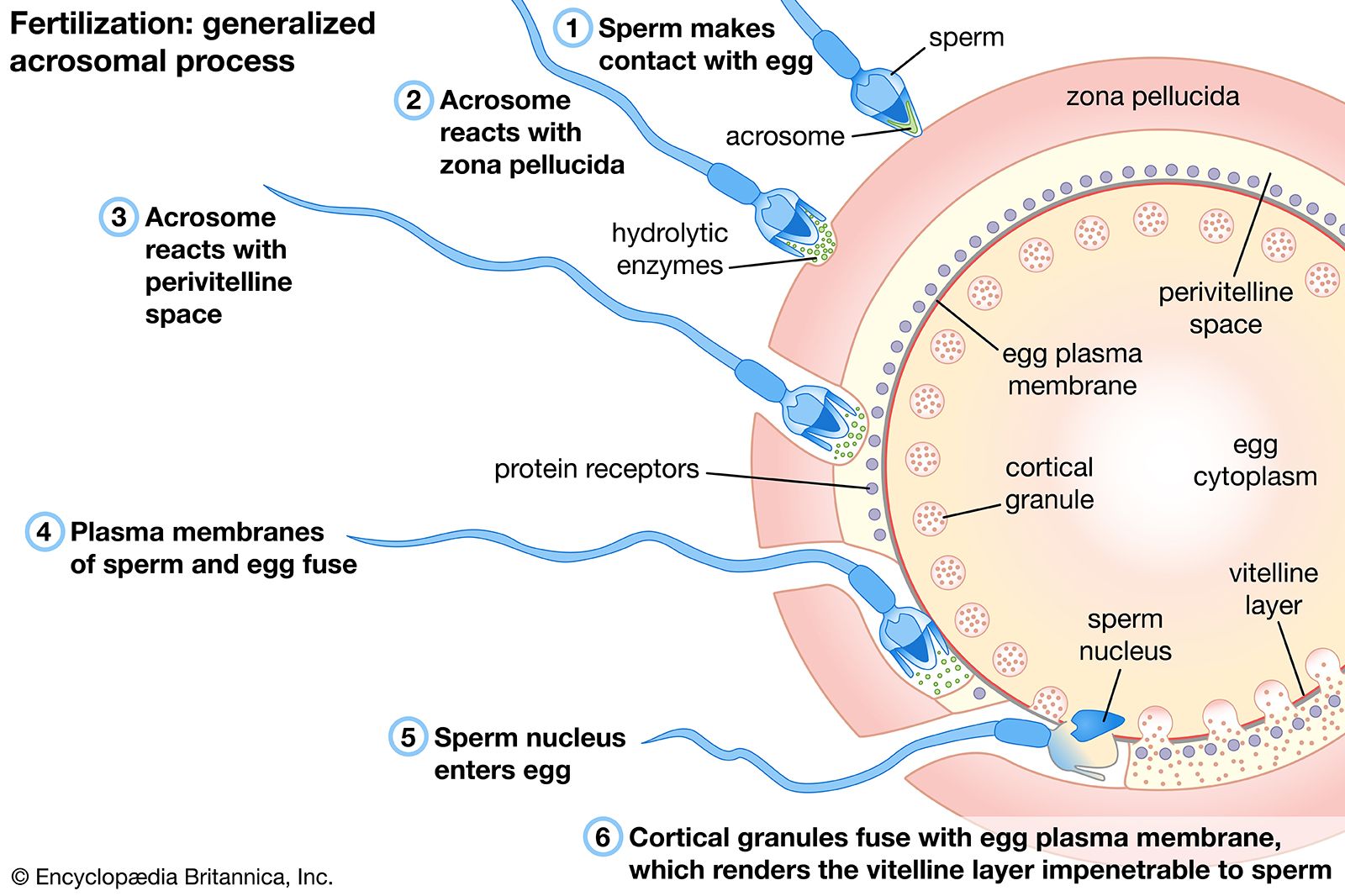


1️⃣ SPERM ACTIVATION & PENETRATION OF CORONA RADIATA
❓ Why activation is needed
- Fresh sperm cannot penetrate the oocyte
- They must first become functionally competent
🔬 Capacitation — the activation step
- Occurs before sperm reach distal uterine tube
- Takes place in:
- Cervix
- Uterine tube
- Mechanism:
- Secretions from cervix & uterine tube remove:
- Glycoprotein
- Cholesterol
- Result: sperm become capable of:
- Acrosomal reaction
- Penetration of ovum
from the acrosomal membrane
🧱 Penetration of corona radiata
- Corona radiata = granulosa cells surrounding secondary oocyte
- Process:
- Viable sperm surround the oocyte
- Undergo acrosomal reaction
- Release hyaluronidase
- Enzyme that breaks down intercellular matrix of corona radiata
- Additional factor:
- Active sperm motility is essential
➡️ Outcome: sperm reach the zona pellucida
2️⃣ ATTACHMENT TO & PENETRATION OF ZONA PELLUCIDA
🧲 Attachment
- Once corona radiata is cleared:
- A sperm binds to zona pellucida
🧪 Acrosomal enzymes released
Enzymes responsible for zona penetration:
- Esterases
- Neuraminidase
- Acrosin
🚫 Zona reaction — block to polyspermy
- Binding of first sperm triggers zona reaction
- What changes?
- Physical properties of zona pellucida are altered
- Prevents attachment of additional sperm
🔥 Underlying mechanism — cortical reaction
- Cortical granules in oocyte release lysosomal enzymes
- Enzymes enter space:
- Between zona pellucida & oocyte cell membrane
- This causes:
- Zona hardening
- Sperm entry blockade
➡️ Outcome: only one sperm proceeds further
3️⃣ FUSION OF SPERM & OOCYTE CELL MEMBRANES
📍 Location
- After zona penetration:
- Sperm enters perivitelline space
🔗 Fusion event
- Sperm head membrane contacts oocyte cell membrane
- Fusion of membranes occurs
- After fusion:
- Cell membranes of sperm & egg break down at contact area
➡️ Outcome: sperm contents enter oocyte cytoplasm
4️⃣ COMPLETION OF MEIOSIS II & PRONUCLEI FORMATION
🧬 Meiosis II completion
- Triggered soon after sperm entry
- Secondary oocyte:
- Completes second meiotic division
- Forms:
- Mature oocyte
- Second polar body
🔵 Pronuclei formation
- Maternal & paternal chromosomes:
- Condense
- Enlarge
→ form female pronucleus and male pronucleus
🧵 Chromosomal preparation
- As pronuclei approach:
- Haploid chromosomes:
- Arrange on a spindle
- Split longitudinally into chromatids
➡️ Outcome: genetic material ready for union
5️⃣ FORMATION OF THE ZYGOTE
🤝 Union
- Male & female pronuclei meet
- Their membranes break down
- Chromosomes intermix
🧠 Definition achieved
- A single diploid cell is formed → zygote
🚀 End of fertilisation
- Fertilisation is now complete
- Zygote prepares for:
- First mitotic division
🧠 ONE-LINE LOGIC SUMMARY (EXAM GOLD)
Capacitation → corona radiata penetration (hyaluronidase + motility) → zona penetration (acrosomal enzymes) → zona reaction (cortical granules) → membrane fusion → meiosis II completion → pronuclei formation → zygote
🧬 WEEK 1, WEEK 2, WEEK 3 —(EXAM GOLD)
WEEK 1 — Cleavage → Morula → Blastocyst → Enters Uterus
1️⃣ Starting Point: The Fertilised Ovum (Zygote)
Logic: What must happen before growth can start?
➡️ Genetic completeness.
- After fertilisation, the ovum has a diploid number of chromosomes (46).
- This is only possible after completion of the second meiotic division of the oocyte.
- Once meiosis II is completed → the cell is now a true zygote.
👉 Only a diploid cell can safely enter mitosis, so cleavage can now begin.
2️⃣ Cleavage: Rapid Cell Division Without Growth
Logic: How do we increase cell number without increasing size?
➡️ Repeated mitosis with cytoplasmic subdivision.
- Cleavage = a series of rapid mitotic divisions.
- Occurs over ~3 days.
- The zygote divides into:
- 2 → 4 → 8 → 16 cells.
- These divisions occur without overall increase in embryo size.
🔑 Key consequences of cleavage:
- Cell number ↑
- Individual cell size ↓
- Total embryo size remains the same
Each resulting cell is called a blastomere.
3️⃣ 16-Cell Stage → Morula
Logic: What do many small cells packed together look like?
➡️ A solid ball.
- Around the 16-cell stage, the embryo becomes a solid sphere of cells.
- This stage is called the morula.
- At this stage:
- No cavity yet
- Cells are tightly packed
🔬 Developmental potential:
- Each blastomere is pluripotential at this stage.
- Can still give rise to multiple tissue types.
4️⃣ Transition: Morula → Blastocyst
Logic: What change allows further differentiation?
➡️ Formation of a cavity.
- Fluid begins to accumulate inside the morula.
- Small spaces coalesce to form a central cavity.
- This cavity is called the blastocoele.
Once this cavity forms, the structure is now called a blastocyst.
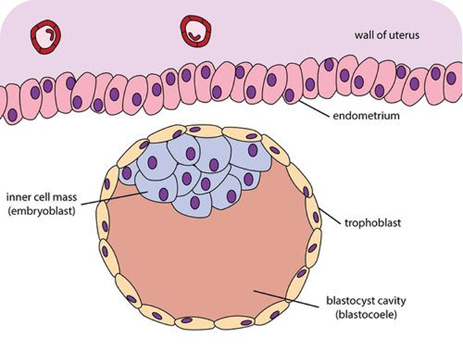
5️⃣ Blastocyst Differentiation: Two Cell Populations
Logic: Cells must now specialize to support implantation and development.
A. Outer Cell Layer → Trophoblast
- The outer cells:
- Flatten
- Thin out to single-cell thickness
- This outer layer becomes the trophoblast.
Role (logic-based):
- Encloses the blastocyst
- Will later participate in implantation and placental formation
B. Inner Cell Mass (Embryoblast)
- The remaining cells do not stay evenly distributed.
- They aggregate at one pole of the blastocyst.
- This cluster is the inner cell mass.
Logic:
- These cells are protected inside
- They will form the embryo proper
6️⃣ Final Structural Summary (End of First Week)
By the end of this sequence, you now have:
- A blastocyst composed of:
- Blastocoele → fluid-filled cavity
- Trophoblast → outer single-cell layer
- Inner cell mass → clustered at one pole
- Cells have begun functional differentiation
- Size remains similar to original zygote, despite many divisions

WEEK 2 — “Week of TWOs”: 2 layers, 2 cavities, 2 membranes, 2 trophoblast layers

🗓️ SECOND WEEK OF DEVELOPMENT
Theme: Implantation + Bilaminar embryonic disc
(“Week of twos” logic applies everywhere)


1️⃣ Implantation & Decidual Reaction (Maternal side)
What is happening?
- Embryo is partly implanted in the endometrium.
Why is this important?
- Implantation triggers decidualisation of uterine stroma.
Decidual reaction (logic):
- Endometrial stromal cells → enlarge + become metabolically active
- These cells:
- Provide nutrition
- Form the maternal component of the placenta
📌 Exam hook:
Placenta = maternal decidua + fetal trophoblast
2️⃣ Trophoblast Differentiation (Fetal invasive system)

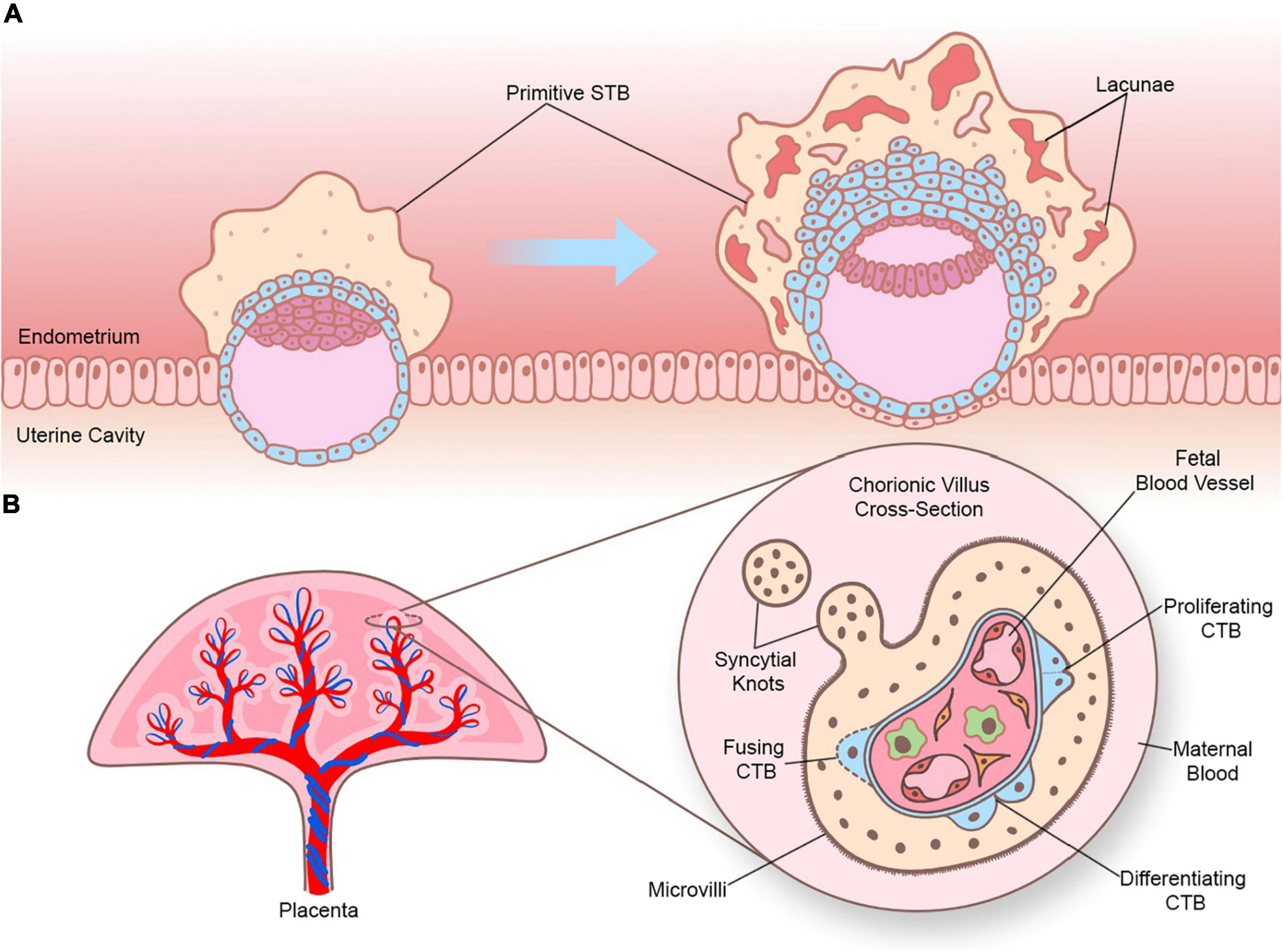
Original trophoblast splits into TWO layers:
A. Cytotrophoblast
- Inner layer
- Single layer of cells
- Mitotically active
- Provides cells for growth of outer layer
B. Syncytiotrophoblast
- Outer layer
- Multinucleated syncytium
- Invasive
- Invades endometrium
- ❌ At this stage: NOT invading endometrial blood vessels
📌 Key timing point:
Invasion of vessels happens later, after lacunae formation.
3️⃣ Inner Cell Mass → Bilaminar Embryonic Disc
Inner cell mass differentiates into TWO layers:
A. Epiblast
- Upper layer
- Columnar cells
- Gives rise to:
- Amniotic cavity
- All three germ layers later
B. Hypoblast
- Lower layer
- Cuboidal cells
- Contributes to:
- Exocoelomic membrane
- Yolk sac lining
Together they form:
➡️ Bilaminar embryonic disc
4️⃣ Formation of Amniotic Cavity (Above epiblast)
Step-by-step logic:
- A cavity develops within epiblast
- This becomes the amniotic cavity
- Some epiblast cells → amnioblasts
- Amnioblasts:
- Line the cavity
- Secrete amniotic fluid
📌 Orientation memory:
- Amniotic cavity = above epiblast
5️⃣ Formation of Primary Yolk Sac (Below hypoblast)


How it forms:
- Hypoblast → forms exocoelomic membrane
- This membrane lines a new cavity
- Cavity = Primary yolk sac
Function:
- Early nutrition
- Supports embryo before placenta is functional
📌 Orientation memory:
- Yolk sac = below hypoblast
6️⃣ Day ~12 Changes – Lacunar Stage Begins


Changes in syncytiotrophoblast:
- Small clefts form → lacunae
- Lacunae:
- Communicate with maternal endometrial sinusoids
- Allow maternal blood to enter
➡️ First uteroplacental circulation begins
📌 Still:
- Syncytiotrophoblast is invasive
- Now functionally nutritive
7️⃣ Extra-Embryonic Coelom (Chorionic Cavity)
How it forms:
- Clefts appear:
- Between exocoelomic membrane
- And cytotrophoblast
- These clefts merge
- Result → extra-embryonic coelom
Effect:
- Almost completely surrounds embryo
- Embryo now suspended inside chorionic cavity
📌 Terminology:
- Extra-embryonic coelom = chorionic cavity
8️⃣ Day ~13 – Chorionic Villi & Structural Organisation
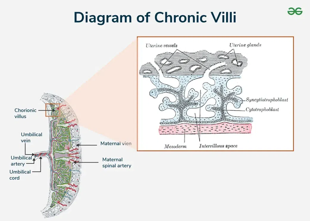
A. Primary Chorionic Villi
- Cytotrophoblast proliferates
- Forms finger-like projections
- Project into lacunae
- These are primary chorionic villi
➡️ First step in placental villous tree
B. Secondary Yolk Sac
- Due to expansion of chorionic cavity:
- Primary yolk sac is reduced
- New cavity forms → secondary yolk sac
C. Embryo Proper Status
- Still bilaminar
- Epiblast + hypoblast remain closely apposed
- Two cavities enlarging:
- Amniotic cavity above
- Yolk sac below

D. Connecting Stalk
- Made of extra-embryonic mesoderm
- Connects embryo to trophoblast
- Becomes:
➡️ Umbilical cord (future)
9️⃣ Completion of Implantation
Final surface event:
- Uterine epithelium reforms
- Conceptus becomes completely embedded
- No surface defect remains
📌 Exam phrase:
“Conceptus fully engulfed by endometrium”
🔟 Hormonal Function – hCG Production
Source:
- Syncytiotrophoblast
Timing:
By end of second week
Actions of hCG:
- Maintains corpus luteum
- Corpus luteum:
- Continues progesterone secretion
- Maintains endometrial thickness
Clinical significance:
- hCG is:
- Secreted into maternal blood
- Excreted in urine
- Basis of early pregnancy test
🧠 FINAL LOGIC LOCK (EXAM-PERFECT SUMMARY)
- Implantation → decidual reaction
- Trophoblast → cytotrophoblast + syncytiotrophoblast
- Inner cell mass → epiblast + hypoblast
- Cavities:
- Amniotic cavity (above epiblast)
- Yolk sac (below hypoblast)
- Lacunae → maternal blood supply
- Extra-embryonic coelom → chorionic cavity
- Primary chorionic villi begin placentation
- Connecting stalk → umbilical cord
- Syncytiotrophoblast → hCG
SECOND WEEK OF DEVELOPMENT — MASTER TABLE (WEEK OF TWOs)
GLOBAL THEME
Concept | Core Idea |
Week identity | “Week of TWOs” |
Major processes | Implantation + bilaminar embryonic disc formation |
Logic pattern | Everything appears in pairs (2 layers, 2 cavities, 2 membranes, 2 trophoblast layers) |
1. IMPLANTATION & DECIDUAL REACTION (MATERNAL SIDE)
Aspect | Details |
Implantation status | Embryo partly implanted in endometrium |
Triggered maternal response | Decidualisation of uterine stroma |
Cellular change | Endometrial stromal cells enlarge + become metabolically active |
Function of decidual cells | Provide nutrition + form maternal component of placenta |
Placenta composition (exam hook) | Maternal decidua + fetal trophoblast |
2. TROPHOBLAST DIFFERENTIATION (FETAL INVASIVE SYSTEM)
Feature | Cytotrophoblast | Syncytiotrophoblast |
Position | Inner layer | Outer layer |
Cell structure | Single layer | Multinucleated syncytium |
Mitotic activity | Mitotically active | Not mitotically active |
Function | Supplies cells to outer layer | Invasive |
Action on endometrium | — | Invades endometrium |
Blood vessel invasion (early) | — | ❌ Not invading vessels yet |
Later role | Forms villi | Nutrition + hCG secretion |
📌 Timing lock:
Vessel invasion occurs after lacunae formation, not initially.
3. INNER CELL MASS → BILAMINAR EMBRYONIC DISC
Feature | Epiblast | Hypoblast |
Position | Upper layer | Lower layer |
Cell type | Columnar cells | Cuboidal cells |
Major derivatives | Amniotic cavity + all 3 germ layers later | Exocoelomic membrane + yolk sac lining |
Structural outcome | - | - |
➡️ Together form: Bilaminar embryonic disc
4. AMNIOTIC CAVITY FORMATION (ABOVE EPIBLAST)
Step | Description |
Initial event | Cavity develops within epiblast |
Cavity name | Amniotic cavity |
Cell differentiation | Some epiblast cells → amnioblasts |
Amnioblast function | Line cavity + secrete amniotic fluid |
Orientation memory | Amniotic cavity = above epiblast |
5. PRIMARY YOLK SAC FORMATION (BELOW HYPOBLAST)
Aspect | Details |
Membrane source | Hypoblast forms exocoelomic membrane |
Cavity lined | Primary yolk sac |
Main function | Early nutrition |
Functional timing | Supports embryo before placenta is functional |
Orientation memory | Yolk sac = below hypoblast |
6. DAY ~12 — LACUNAR STAGE (FUNCTIONAL TURNING POINT)
Feature | Description |
Structure affected | Syncytiotrophoblast |
Change | Small clefts form → lacunae |
Lacunae connection | Communicate with maternal endometrial sinusoids |
Blood entry | Maternal blood enters lacunae |
Functional result | First uteroplacental circulation begins |
Syncytiotrophoblast status | Still invasive, now nutritive |
7. EXTRA-EMBRYONIC COELOM (CHORIONIC CAVITY)

Step | Description |
Initial change | Clefts appear between exocoelomic membrane & cytotrophoblast |
Progression | Clefts merge |
Final cavity | Extra-embryonic coelom |
Alternate name | Chorionic cavity |
Effect on embryo | Embryo almost completely surrounded |
Suspension | Embryo suspended within chorionic cavity |
8. DAY ~13 — STRUCTURAL ORGANISATION & PLACENTAL PRIMORDIA
A. PRIMARY CHORIONIC VILLI
Feature | Details |
Cellular event | Cytotrophoblast proliferates |
Morphology | Finger-like projections |
Direction | Project into lacunae |
Name | Primary chorionic villi |
Significance | First step in placental villous tree |
B. SECONDARY YOLK SAC
Feature | Details |
Cause | Expansion of chorionic cavity |
Effect | Primary yolk sac reduced |
New structure | Secondary yolk sac |
C. EMBRYO PROPER STATUS
Feature | Status |
Disc type | Still bilaminar |
Germ layers | Epiblast + hypoblast closely apposed |
Cavities | Amniotic cavity above + yolk sac below |
D. CONNECTING STALK
Feature | Details |
Composition | Extra-embryonic mesoderm |
Function | Connects embryo to trophoblast |
Future derivative | Umbilical cord |
9. COMPLETION OF IMPLANTATION
Feature | Description |
Surface event | Uterine epithelium reforms |
Conceptus status | Completely embedded |
Surface defect | None |
Exam phrase | “Conceptus fully engulfed by endometrium” |
10. HORMONAL FUNCTION — hCG
Aspect | Details |
Source | Syncytiotrophoblast |
Time of secretion | By end of second week |
Primary action | Maintains corpus luteum |
Corpus luteum effect | Continues progesterone secretion |
Endometrial role | Maintains thickness |
Clinical relevance | Basis of early pregnancy test |
Detection | Present in maternal blood & urine |
FINAL EXAM LOGIC LOCK (ONE-GLANCE)
Domain | Key Pair |
Implantation | Decidual reaction |
Trophoblast | Cyto + Syncytio |
Embryonic disc | Epiblast + Hypoblast |
Cavities | Amniotic (up) + Yolk sac (down) |
Nutrition | Lacunae + maternal blood |
Surrounding space | Extra-embryonic coelom |
Placenta start | Primary chorionic villi |
Connection | Connecting stalk → umbilical cord |
Hormone | Syncytiotrophoblast → hCG |
WEEK 3 — Gastrulation → Trilaminar Disc (ECTO • MESO • ENDO)

(The moment the embryo becomes “organ-capable”)


1️⃣ Starting Point: What Exists at the End of Week 2?
Logical setup
Before Week 3 begins, the embryo is simple and flat, but organized.
Structures present
- Bilaminar embryonic disc
- Epiblast (upper layer)
- Hypoblast (lower layer)
- These two layers:
- Are closely apposed
- Form two elliptical plates
- Together = bilaminar embryonic disc
🧠 Key logic
All further development comes from rearranging and expanding what already exists — not adding something foreign.
2️⃣ The Core Event of Week 3: Gastrulation
Definition (must-know)
Gastrulation = the process by which the embryo forms three germ layers.
Why gastrulation is critical
- Converts a two-layered disc → three-layered disc
- Establishes the basic body plan
- Makes organ development possible
🧠 Exam anchor
“No gastrulation → no organs.”
3️⃣ Renaming of Existing Layers (Conceptual Shift)
Logical transformation
Once gastrulation begins, the original two layers get new identities based on their final roles.
Before | After gastrulation |
Epiblast | Ectoderm |
Hypoblast | does not become Endoderm |
📌 Important clarification:
- Epiblast ≠ disappears
- It becomes ectoderm and also produces mesoderm
🧠 Key rule
All three germ layers originate from the epiblast.
4️⃣ Formation of the Third Layer: Intra-embryonic Mesoderm
How the third layer appears (logic, not memorization)
- Cells from the ectoderm (epiblast) migrate inward
- These migrating cells settle between ectoderm and endoderm
- This new middle layer is the intra-embryonic mesoderm
📌 Positioning:
- Ectoderm → outer
- Mesoderm → middle
- Endoderm → inner
🧠 Why this matters
Without a middle layer, you cannot build strength, movement, or support.
5️⃣ The Trilaminar Embryonic Disc (End Result)
Final configuration
After gastrulation, the embryo is a:
👉 Trilaminar embryonic disc, composed of:
- Ectoderm
- Mesoderm
- Endoderm
This is the basic structural blueprint for the entire human body.
6️⃣ Functional Logic of the Three Germ Layers
(“Outer skin, middle strength, inner lining”)
🟦 ECTODERM — “Outer skin & control”
- Forms:
- Epidermis (skin covering)
- Nervous system
- Think:
- Protection
- Sensation
- Communication
🧠 Memory hook:
Ecto = external + electrical (nervous system)
🟥 MESODERM — “Support, movement, circulation”
- Forms:
- Skeletal tissue
- Connective tissue
- Muscle
- Think:
- Strength
- Framework
- Motion
🧠 Memory hook:
Meso = middle + mechanical
🟩 ENDODERM — “Inner lining & exchange”
- Forms:
- Gastrointestinal tract lining
- Respiratory tract lining
- Think:
- Absorption
- Secretion
- Gas exchange
🧠 Memory hook:
Endo = inside
7️⃣ High-Yield Generalisation (Exam Gold)
One-line logic summary
- Ectoderm → covering + nervous system
- Mesoderm → skeletal, connective & muscle tissues
- Endoderm → GI & respiratory linings
📌 This generalisation is explicitly stated and commonly examined.
8️⃣ Why Week 3 Is a Turning Point (Conceptual Closure)
- Before Week 3 → embryo is layered but not functional
- After Week 3 → embryo has:
- Direction
- Identity
- Organ-forming capacity
🧠 Final lock
Week 3 = the embryo commits to becoming human in structure.
DEVELOPMENT OF ECTODERM & MESODERM
1. Primitive Streak Formation (Start of Gastrulation)


Logical trigger
- End of Week 2 → bilaminar disc exists (epiblast + hypoblast)
- A midline groove appears at the caudal end → primitive streak
Step-by-step logic
- Primitive streak appears
- Groove-like midline depression
- Marks beginning of gastrulation
- Week 3
- Streak deepens
- Primitive node forms at cephalic end of streak
- Cell migration
- Ectodermal (epiblast) cells migrate towards the streak
- Cells detach, move beneath ectoderm, and spread laterally
- Result
- Formation of intra-embryonic mesoderm
- Disc becomes trilaminar
Critical exceptions (areas WITHOUT mesoderm)
- Prochordal plate (cephalic)
- Cloacal plate (caudal)
Fate of these regions
- Prochordal plate
- Replaced by buccopharyngeal membrane
- Temporarily seals future oral cavity
- Week 4 → membrane breaks → communication between gut tube & amniotic cavity
- Cloacal plate
- Replaced by cloacal membrane
2. Notochord Formation (Midline Axis Builder)


Logical sequence
- Origin
- Cells from primitive node
- Migration
- Move cranially toward buccopharyngeal membrane
- Intermediate structure
- Notochordal plate forms
- Final structure
- Plate folds inward → solid notochord
Functional significance (must-know)
- Lies beneath future neural tube
- Establishes:
- Longitudinal axis of embryo
- Nucleus pulposus of intervertebral discs
3. Neurulation (Brain & Spinal Cord Formation)
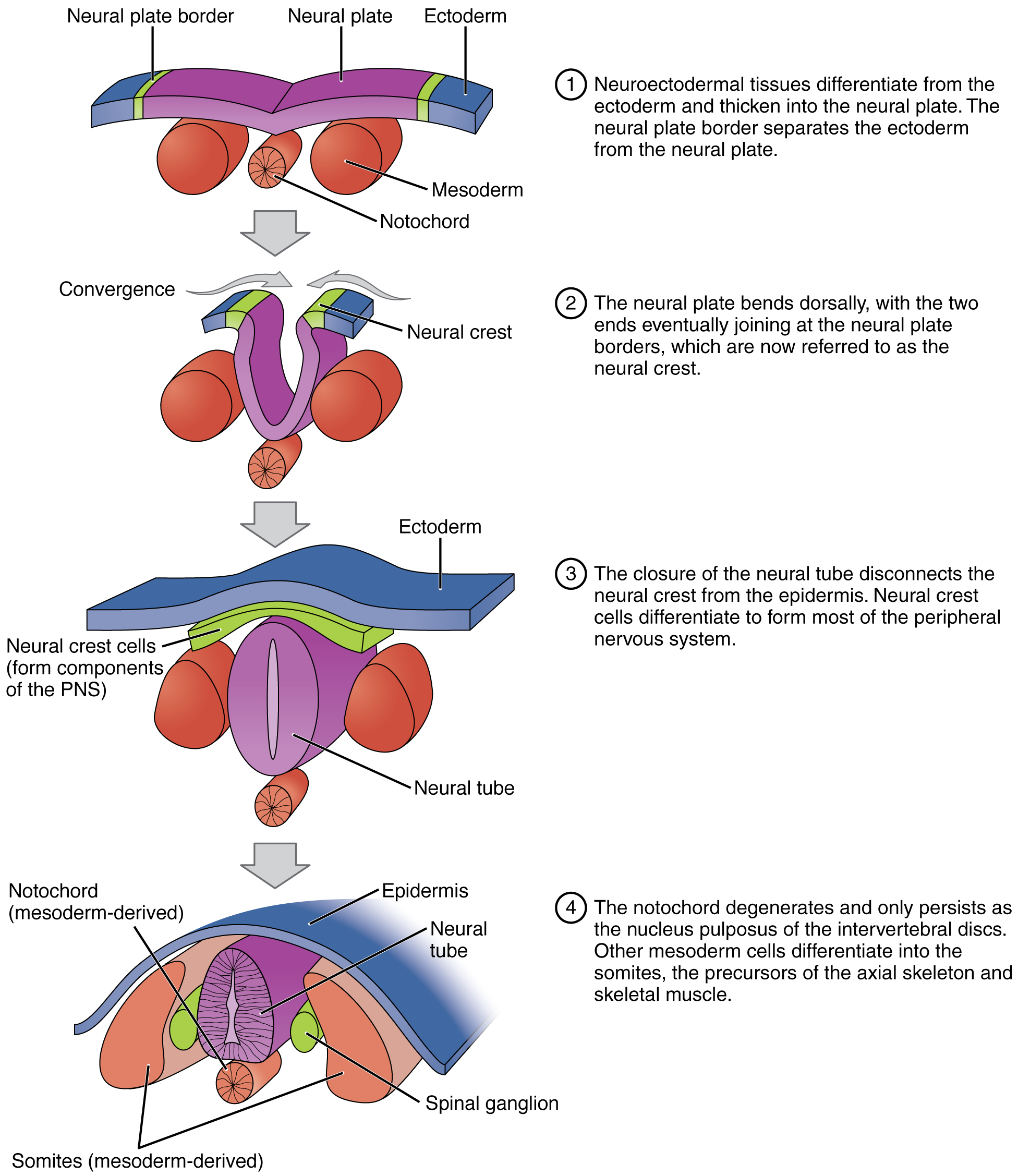


Definition
- Neurulation = formation of brain and spinal cord
Induction logic
- Day ~19
- Notochord + underlying mesoderm induce ectoderm
- Ectoderm → Neuroectoderm
- Forms neural plate
Morphological sequence
- Neural plate
- Appears at cranial end first
- Day 20
- Mid-region: narrow
- Caudal end: expanded
- Neural groove
- Plate deepens
- Neural tube
- Groove closes
Neuropores
- Anterior (cranial) neuropore
- Posterior (caudal) neuropore
- Initially open → later close
Neural crest cells
- Form at junction of neuroectoderm & surface ectoderm
- Detach before tube closure
- Form discrete migrating cell populations
Neural crest derivatives (full list)
- Dorsal root ganglia
- Cranial nerve ganglia
- Enteric ganglia
- Autonomic ganglia
- Connective tissue of face
- Bones of skull
- Adrenal medulla
- Glial cells
- Schwann cells
- Melanocytes
- Parts of meninges
- Parts of teeth
4. Further Development of the Mesoderm
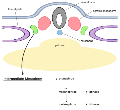
Spatial organisation (Day ~17)
- Mesoderm thickest near midline → Paraxial mesoderm
- Moving laterally:
- Intermediate mesoderm
- Lateral plate mesoderm
Lateral plate changes (Day ~19)
- Clefts appear
- Plate splits into two layers:
- Parietal (somatic) layer
- Covers amniotic sac
- Visceral (splanchnic) layer
- Covers yolk sac
Coelom formation
- Clefts merge → Intra-embryonic coelom
- Precursor of:
- Pericardial cavity
- Pleural cavities
- Peritoneal cavity
- Intra- and extra-embryonic coeloms are continuous
Intermediate mesoderm
- Lies between paraxial & lateral plate
- Gives rise to urogenital system

5. Segmentation of the Mesoderm – Paraxial Mesoderm

Somite formation
- Paired blocks along craniocaudal axis
- First pair: ~Day 20
- Rate: ~3 pairs/day
- Total: 42–44 pairs (not all persist)
Clinical logic
- Embryo age correlates with somite number
🧬 WEEK 3: GASTRULATION → TRILAMINAR DISC (MASTER TABLE SET)
TABLE 1 — Starting Point (End of Week 2)
Aspect | Details |
Embryonic stage | Bilaminar embryonic disc |
Layers present | Epiblast (upper) + Hypoblast (lower) |
Shape | Two closely apposed elliptical plates |
Complexity | Simple, flat, organized but not organ-capable |
Core logic | All future structures arise by rearrangement and migration, not new material |
TABLE 2 — Gastrulation (Core Event of Week 3)
Feature | Description |
Definition | Formation of three germ layers |
Structural change | Bilaminar → Trilaminar disc |
Functional impact | Establishes basic body plan |
Developmental significance | Makes organ development possible |
Exam anchor | “No gastrulation → no organs” |
TABLE 3 — Fate of Original Layers (Conceptual Shift)
Original Layer | Post-Gastrulation Fate | Key Clarification |
Epiblast | Becomes ectoderm AND gives rise to mesoderm + endoderm | All 3 germ layers originate from epiblast |
Hypoblast | Does NOT become endoderm | Replaced by migrating epiblast cells |
TABLE 4 — Formation of Intra-Embryonic Mesoderm
Step | Description |
Cell source | Epiblast (future ectoderm) |
Movement | Cells migrate towards primitive streak, then inward |
Final position | Between ectoderm & endoderm |
Result | Formation of intra-embryonic mesoderm |
Positional order | Ectoderm (outer) → Mesoderm (middle) → Endoderm (inner) |
Logic | Middle layer essential for strength, movement, support |
TABLE 5 — Trilaminar Embryonic Disc (End Result)
Germ Layer | Position | Core Role |
Ectoderm | Outer | Covering + control |
Mesoderm | Middle | Support + movement |
Endoderm | Inner | Lining + exchange |
Developmental status | Embryo now organ-capable |
TABLE 6 — Functional Logic of Germ Layers (Exam Gold)
Germ Layer | Major Derivatives | Memory Hook |
Ectoderm | Epidermis, nervous system | External + electrical |
Mesoderm | Skeletal tissue, connective tissue, muscle | Middle + mechanical |
Endoderm | GI lining, respiratory lining | Inside |
TABLE 7 — Primitive Streak (Start of Gastrulation)
Feature | Details |
Time | End of Week 2 → Week 3 |
Location | Midline, caudal end of disc |
Structure | Groove-like depression |
Associated structure | Primitive node at cephalic end |
Function | Entry point for cell migration |
Outcome | Formation of intra-embryonic mesoderm |
TABLE 8 — Cell Migration via Primitive Streak
Step | Event |
1 | Epiblast cells migrate toward streak |
2 | Cells detach and move inward |
3 | Spread laterally beneath ectoderm |
4 | Form mesoderm |
Final result | Trilaminar disc |
TABLE 9 — Regions WITHOUT Mesoderm (Critical Exceptions)
Region | Location | Fate |
Prochordal plate | Cephalic | Becomes buccopharyngeal membrane |
Cloacal plate | Caudal | Becomes cloacal membrane |
TABLE 10 — Buccopharyngeal & Cloacal Membranes
Feature | Buccopharyngeal | Cloacal |
Origin | Prochordal plate | Cloacal plate |
Function | Temporarily seals oral cavity | Seals caudal opening |
Fate | Breaks in Week 4 | Persists longer |
Result after rupture | Communication between gut tube & amniotic cavity | Future anal opening |
TABLE 11 — Notochord Formation
Stage | Description |
Origin | Primitive node |
Migration | Cranial toward buccopharyngeal membrane |
Intermediate | Notochordal plate |
Final structure | Solid notochord |
Position | Beneath future neural tube |
TABLE 12 — Notochord: Functions
Function | Significance |
Establishes | Longitudinal axis |
Induces | Neural plate formation |
Adult remnant | Nucleus pulposus of intervertebral discs |
TABLE 13 — Neurulation (Brain & Spinal Cord Formation)
Aspect | Details |
Definition | Formation of brain + spinal cord |
Time | Starts ~Day 19 |
Induction | Notochord + mesoderm induce ectoderm |
Ectoderm becomes | Neuroectoderm |
Initial structure | Neural plate |
TABLE 14 — Morphological Sequence of Neurulation
Stage | Description |
Neural plate | Appears first cranially |
Neural groove | Plate deepens |
Neural tube | Groove closes |
Neuropores | Anterior & posterior initially open |
Closure | Both neuropores close later |
TABLE 15 — Neural Crest Cells
Feature | Details |
Origin | Junction of neuroectoderm & surface ectoderm |
Timing | Detach before neural tube closure |
Nature | Highly migratory cell population |
TABLE 16 — Neural Crest Derivatives (Complete List)
Category | Derivatives |
Nervous system | Dorsal root ganglia, cranial nerve ganglia, autonomic & enteric ganglia |
Support cells | Schwann cells, glial cells |
Endocrine | Adrenal medulla |
Pigmentation | Melanocytes |
Craniofacial | Bones of skull, connective tissue of face |
Meninges | Parts of meninges |
Dental | Parts of teeth |
TABLE 17 — Mesoderm: Spatial Organisation (~Day 17)
Region | Position | Major Outcome |
Paraxial mesoderm | Adjacent to midline | Somites |
Intermediate mesoderm | Between paraxial & lateral plate | Urogenital system |
Lateral plate mesoderm | Most lateral | Body cavities |
TABLE 18 — Lateral Plate Mesoderm Changes (~Day 19)
Step | Event |
1 | Clefts appear |
2 | Plate splits |
Layers formed | Parietal (somatic) + Visceral (splanchnic) |
Parietal layer covers | Amniotic sac |
Visceral layer covers | Yolk sac |
TABLE 19 — Intra-Embryonic Coelom
Feature | Description |
Formation | Fusion of clefts |
Becomes | Pericardial, pleural, peritoneal cavities |
Continuity | Continuous with extra-embryonic coelom |
TABLE 20 — Paraxial Mesoderm Segmentation (Somites)
Feature | Detail |
First appearance | Day ~20 |
Formation rate | ~3 pairs/day |
Total number | 42–44 pairs |
Arrangement | Paired blocks along craniocaudal axis |
Clinical use | Embryo age estimation |
Week 4 differentiation
- Dermomyotome
- Connective tissue
- Skeletal muscle
- Sclerotome
- Bone
- Cartilage
Vertebral column
- Sclerotomal cells surround:
- Notochord
- Spinal cord
6. Somite Development (Detailed Fate Mapping)
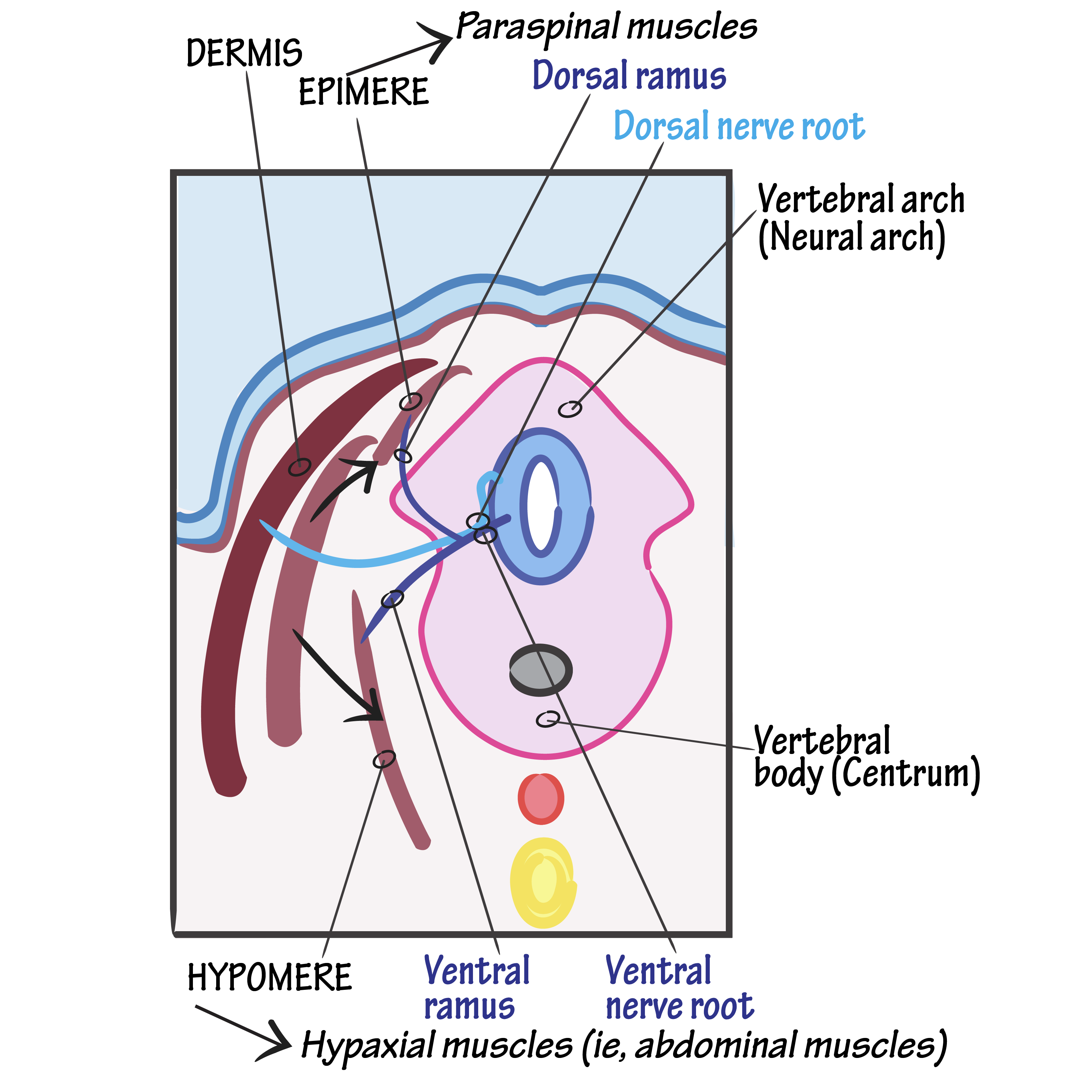

Cellular rearrangement
- Medial mesenchymal cells
- → Sclerotomes
- Ventrolateral cells
- → Myotomes
- Remaining cells
- → Dermatomes
Myotome subdivision
- Dorsal epimeres
- → Epaxial muscles
- (Erector spinae)
- Ventral hypomeres
- → Hypaxial muscles
- Body wall muscles
Limb muscles
- Ventrolateral somite cells in limb regions migrate
- Form limb musculature
Dermatome fate
- Form dermis
- Lie beneath epidermis (ectodermal)
Key neurological principle
- Migrating myotomes & dermatomes carry their original segmental innervation
- Explains dermatomes & myotomes in adults
7. Lateral Plate Mesoderm (Serous Cavities & Gut Wall)


Structural arrangement
- Two layers enclosing intra-embryonic coelom
Differentiation
- lateral plate Mesoderm becomes thin sheets → serous membranes
Layers and names
- Parietal layer
- Lines future body wall
- Also called somatopleure
- Visceral layer
- Covers endodermal gut tube
- Also called splanchnopleure
Structures formed
- Pleura
- Pericardium
- Peritoneum
- Smooth muscle of gut
- Connective tissue of gut wall
Final Big-Picture Logic Lock 🔒
- Primitive streak → mesoderm
- Primitive node → notochord
- Notochord → neural induction
- Paraxial mesoderm → somites
- Somites → bone, muscle, dermis
- Lateral plate → body cavities & gut coverings
- Neural crest → wide multi-system derivatives
🧬 ENDODERM + FOLDING (WEEK 4)
1. Endoderm – What it forms (FOUNDATION)
Core principle
- Endoderm = internal epithelial linings + glandular parenchyma
- It forms structures that deal with absorption, secretion, and internal exchange.
Exact derivatives (no omissions)
The endoderm gives rise to:
- Epithelial lining of the gastrointestinal (GI) tract
- Epithelial lining of the respiratory tract
- Parenchymal (functional) cells of:
- Liver
- Pancreas
- Thyroid gland
- Parathyroid glands
- Epithelial lining of the urinary bladder
📌 Key logic
Endoderm → lining + secretory tissue
Mesoderm → muscle/connective tissue around these linings
Ectoderm → external surface + nervous system
2. Why folding is necessary (TRANSITION LOGIC)
Initial problem
- Early embryo is a flat trilaminar disc
- GI tract must become a tube
- Flat structures cannot enclose organs
Solution
➡️ The embryo undergoes folding in two planes to convert a flat sheet into a 3D body with a gut tube
3. Fourth Week – Folding of the Embryo (TIMING)
- Occurs in the 4th week
- Folding happens in two directions:
- Longitudinal (cephalocaudal) folding
- Lateral (transverse) folding
These processes occur together, not separately.
4. Longitudinal (Cephalocaudal) Folding – WHY & HOW
Primary cause
- Rapid enlargement of the cranial neural tube
- This forms the brain
- Brain growth is disproportionate → forces bending
Timing
- Occurs between day 21 and day 24
Mechanical result
- Embryo bends so that:
- Head and tail move toward each other
- Flat disc becomes curved
5. Effect of Longitudinal Folding on Endoderm (KEY OUTCOME)
Initial state
- Endoderm is a flat sheet
- It has a wide communication with the yolk sac
During folding
- Endoderm rolls inward → forms a tube-like structure
- This tube is the primitive gut tube
- Connection to yolk sac becomes:
- Progressively narrower
- Due to increasing folding
📌 Critical logic
- Folding → inward movement of endoderm
- Inward movement → tube formation
- Tube formation → future GI tract
6. Role of the Amniotic Cavity in Folding (MECHANICAL DRIVER)
What the amniotic cavity does
- Expands rapidly
- Pushes inward at:
- Cranial end
- Caudal end
Result
- Enhances:
- Head fold
- Tail fold
- Increases the degree of longitudinal bending
7. Vitello-intestinal (Vitelline) Duct Formation
Initial connection
- Gut tube ↔ yolk sac via a wide opening
During folding
- Amniotic cavity pinches this connection
- The opening narrows to form:
- Vitello-intestinal (vitelline) duct
Fate
- This duct is temporary
- Later disappears completely
📌 Exam logic
Wide connection → narrowing → duct → disappearance
8. Lateral (Transverse) Folding – WHY & EFFECT
Primary cause
- Enlargement of the somites
- Somites grow laterally and ventrally
Result
- Embryo folds from left and right sides toward the midline
- This:
- Completes enclosure of the gut tube
- Closes the ventral body wall (except umbilical region)
9. Yolk Sac – Role and Fate
Early role
- Provides early nutrition to the embryo
After first month
- Nutritional role is lost
- Yolk sac becomes:
- Vestigial
- Lies freely in the chorionic cavity
📌 Important distinction
- Yolk sac is not placental nutrition
- It is an early, temporary support structure
10. Final Integrated Logic Chain (ONE-FLOW SUMMARY)
- Endoderm forms internal epithelial linings and glandular parenchyma
- Embryo starts as a flat disc → cannot house organs
- Rapid brain growth + somite enlargement → forces folding
- Folding occurs longitudinally and laterally in week 4
- Endoderm rolls inward → forms gut tube
- Wide yolk sac connection narrows → vitelline duct
- Vitelline duct later disappears
- Yolk sac loses function → becomes vestigial
🧬 TABLE 1: ENDODERM — CORE PRINCIPLE & DERIVATIVES
Aspect | Details (EXACT, NO OMISSION) |
Core definition | Endoderm forms internal epithelial linings + glandular parenchyma |
Functional theme | Structures involved in absorption, secretion, internal exchange |
GI tract | Epithelial lining of entire gastrointestinal tract |
Respiratory tract | Epithelial lining of respiratory tract |
Glandular parenchyma | Liver, Pancreas, Thyroid gland, Parathyroid glands |
Urinary system | Epithelial lining of urinary bladder |
What endoderm does NOT form | Muscle, connective tissue (mesoderm) ; nervous system & skin (ectoderm) |
🧠 TABLE 2: GERM LAYER LOGIC (EXAM INTEGRATION)
Germ Layer | Primary Contribution |
Endoderm | Internal epithelial linings + secretory parenchyma |
Mesoderm | Muscle, connective tissue, blood vessels, supporting framework |
Ectoderm | External surface epithelium + nervous system |
🔄 TABLE 3: WHY EMBRYONIC FOLDING IS NECESSARY
Problem | Reason |
Initial embryo shape | Flat trilaminar disc |
Limitation | Flat structure cannot enclose organs |
GI tract requirement | Must become a tube |
Solution | Folding in two planes to form a 3D body |
⏱️ TABLE 4: TIMING & PLANES OF FOLDING (WEEK 4)
Feature | Details |
Week of occurrence | 4th week of development |
Planes of folding | Longitudinal (cephalocaudal) + Lateral (transverse) |
Relationship | Occur simultaneously, not sequentially |
🔁 TABLE 5: LONGITUDINAL (CEPHALOCAUDAL) FOLDING — CAUSE & MECHANICS
Aspect | Details |
Primary cause | Rapid enlargement of cranial neural tube (brain growth) |
Growth pattern | Disproportionate cranial growth |
Timing | Day 21–24 |
Mechanical effect | Head and tail bend toward each other |
Shape change | Flat disc → curved embryo |
🧪 TABLE 6: EFFECT OF LONGITUDINAL FOLDING ON ENDODERM
Stage | Endoderm Status |
Initial | Flat sheet with wide yolk sac communication |
During folding | Rolls inward |
Structural result | Primitive gut tube formation |
Yolk sac connection | Becomes progressively narrower |
Final logic | Folding → inward movement → tube → future GI tract |
🌊 TABLE 7: ROLE OF AMNIOTIC CAVITY IN FOLDING
Aspect | Effect |
Growth pattern | Rapid expansion |
Direction of force | Inward pressure at cranial + caudal ends |
Folding enhanced | Head fold + tail fold |
Net effect | Increased longitudinal bending |
🔗 TABLE 8: VITELLO-INTESTINAL (VITELLINE) DUCT FORMATION
Stage | Description |
Initial state | Gut tube ↔ yolk sac via wide opening |
During folding | Amniotic cavity pinches the connection |
Intermediate form | Vitello-intestinal (vitelline) duct |
Fate | Temporary structure → disappears completely |
Exam sequence | Wide opening → narrowing → duct → disappearance |
↔️ TABLE 9: LATERAL (TRANSVERSE) FOLDING — CAUSE & EFFECT
Aspect | Details |
Primary cause | Enlargement of somites |
Direction of growth | Laterally and ventrally |
Folding movement | Left + right sides move toward midline |
Major outcomes | • Complete enclosure of gut tube• Ventral body wall closure |
Exception | Umbilical region remains open |
🥚 TABLE 10: YOLK SAC — ROLE & FATE
Phase | Details |
Early role | Provides early embryonic nutrition |
After 1st month | Nutritional role lost |
Final status | Vestigial structure |
Location later | Lies freely in chorionic cavity |
Important distinction | Not placental nutrition |
🔗 TABLE 11: FINAL INTEGRATED LOGIC CHAIN (EXAM FLOW)
Step | Event |
1 | Endoderm forms internal epithelial linings + glands |
2 | Embryo starts as flat trilaminar disc |
3 | Brain growth + somite enlargement force folding |
4 | Folding occurs longitudinally + laterally in week 4 |
5 | Endoderm rolls inward → gut tube |
6 | Yolk sac connection narrows |
7 | Vitelline duct forms then disappears |
8 | Yolk sac becomes vestigial |